


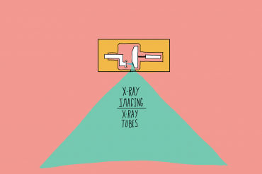
A Medical Student’s Guide to: X-Ray Imaging – The X-Ray Tube
Online Tutoring
I offer online tuition to students studying Biology, Chemistry and Physics at secondary and tertiary levels. I am happy to take on clients from around the world and have a proven track record of top marks achieved by my students. I also tutor for the medical entrance exams, mainly focusing on the UCAT and UCAT ANZ.
To enquire further and set up a free consultation, please use the contact form on this website, drop me an email or contact me through SuperProf.
How do we produce X-Rays?


We create X-Rays in a device called an X-Ray tube. For most medical students, all you need to know is that a beam of electrons is fired at a tungsten (most commonly) target, producing a beam of X-Ray photons. Essentially electrical energy is converted to an electromagnetic wave. Unfortunately for us Imaging Sciences students, we need to know a little more than this.
Below is a schematic of an X-Ray tube. It has a lot of parts, but we’ll talk through them.


First, the fundamentals of the X-Ray tube:
The Cathode
- The left of the image contains the cathode and filament. There is also a focussing cup.
- The cathode is the negative side/end of an electrical circuit.
- The filament, normally made from tungsten, has a very high current passed through it, heating the filament.
- When Tungsten is heated, it will emit electrons by thermionic emission.
- There electrons are accelerated across the X-Ray tube towards the target.
- The focussing cup is negatively charged, repulsing the negatively charged electrons. The focussing cup helps “aim” the beam of electrons.
The Anode
- The right of the image contains the anode, the target and a motor.
- The anode is the positively charged end/side of an electrical circuit. Electrons emitted from the cathode accelerate towards the anode across the X-Ray tube.
- The target is commonly made from tungsten (90%) and rhenium (10%). Tungsten has a high atomic number, making it an efficient emitter of X-Rays. It has a high melting point, able to withstand the massive heat generated. Rhenium allows the material to better conduct heat away from the source, resisting damage over time.
- When electrons bombard the tungsten target, X-rays are produced via the Bremsstrahlung reaction (more on this process below).
- The target is connected to a motor by a molybdenum rod. This helps conduct heat away from the focal spot. The motor spins the target rapidly, to reduce the heat produced per area.
- The target is angled slightly (7-15⁰) which creates a thin beam of X-Rays for imaging.
The entire tube is encased in a glass envelope. Within this, the components are kept under a vacuum.
The Bremsstrahlung Effect
The accelerated electrons can interact with the tungsten target in one of three ways.
- They can interact with other electron in the electron sea and generate heat (in the form of infrared radiation).
- They can interact with the nucleus of atoms, emitting X-Ray radiation through braking (Bremsstrahlung emission).
- They can remove inner shell electrons from the atoms, causing outer shell electrons to fill the vacancies and emit characteristic X-Rays.
Bremsstrahlung radiation is created when an electron passed close to a nucleus. The nucleus deflects the electron, changing the direction of travel and applying a deceleration. This deceleration of a charged particle emits photons, including X-Rays. The energy of the photons is continuous, from zero (with zero deceleration) up to the energy of the photon (with total deceleration).


Characteristic X-Rays are created when a vacancy is created in the inner shell (K shell) of an atom’s electron ring. An electron from an outer ring will fall to fill this space and emit a photon of with energy equal to the energy difference between the two rings (Kα, Kβ, Kγ).


These X-Ray photons all combine to create the X-Ray tube’s emission spectrum. The sharp peaks are the characteristic radiation energies, with the continuous line showing all possible Bremsstrahlung energies. The low energy photons are removed from the beam with the use of a filter. This consists of a thin (millimetres) metal plate between the tube and the patient. As photon interaction with matter, and subsequently it’s absorption when travelling through a medium, is inversely proportional to the energy of the photon. Low energy photons interact with matter more, so are absorbed when passing thought the metal sheet. High energy electrons interact less with matter, so pass through the metal sheet. This creates the recognisable tungsten X-Ray tube filtered emission spectrum.


This is a continuation of my A Medical Student’s Guide To: series, and the first in the x-ray technologies posts. Checkout the others below. you can also access my guides to Ultrasound: The Basics and Ultrasound: Imaging Modes.
I always love to connect with my readers. So please do get in touch on Twitter and Linkedin! You can also use the contact form on the website or leave a comment below. You can SUBSCRIBE for updates and post notifications at the bottom of this page.


Comments
Great read!!! Thanks for sharing such a great blog, blog like these are really helpful.