


A Medical Student’s Guide to: Ultrasound Imaging – The Basics
Online Tutoring
I offer online tuition to students studying Biology, Chemistry and Physics at secondary and tertiary levels. I am happy to take on clients from around the world and have a proven track record of top marks achieved by my students. I also tutor for the medical entrance exams, mainly focusing on the UCAT and UCAT ANZ.
To enquire further and set up a free consultation, please use the contact form on this website, drop me an email or contact me through SuperProf.


The Short History of Ultrasound
Ultrasound imaging is a rapidly evolving imaging modality, invaluable in the fields of trauma, general surgery and obstetrics and gynaecology among others. However, the science behind ultrasound, sonography, has been around for over 200 years.
The Italian Biologist, Lazzaro Spallanzani (1729-1799) is often credited with the discovery of ultrasound. He brought two flying nocturnal predators (owls and bats) into his laboratory and studied their behaviour in different light conditions. He observed that the owls refused to fly around his lab when he eliminated all sources of light. However, bats would fly around the room in the dark, even avoiding small bells and chimes suspended from the ceiling on wires. Lazzaro tried blinding the bats with a “red-hot needle” and observed that even the mutilated bats avoided the obstacles. By inserting brass tubes into the bats’ ears, he caused the animals to lose their ability to echolocate and fly into the wires. Spallanzani concluded that the animals used sound to navigate but did not know that the bats were creating the sound themselves.
Following the sinking of the Titanic in 1912, Physicist Paul Langevin invented the worlds first devise for detecting objects underwater using sound. He called his devise the Hydrophone. We now know of this technology as sonar. Langevin’s Hydrophone was the world’s first transducer.
In the 1920s-1940s, ultrasound was used for a myriad of purposes, including physiotherapy, pain relief and sterilisation.
Research into sonography continued, until in 1942 Karl Dussik pioneered the first diagnostic ultrasound tool. An ultrasound beam was used to detect brain tumours through a patient’s skull.
What is Ultrasound?

Here comes the physics bit…
Ultrasound is defined as:
“Sound waves with a frequency higher than what is audible to the human ear”
These are sound waves with a frequency above 20,000Hz (20 kHz).
Waves are a transmission of energy, kinetic in the example of sound waves, through matter. Sound waves are mechanical, longitudinal waves. That is, the direction of oscillation is parallel to the direction the wave is moving. Bands of high pressure move forward through a medium, while the individual particles oscillate. These high pressure ‘peaks’ are called ‘wavefronts’. The waves propagate perpendicular to the wavefronts.
Wave properties:
- Wavelength – Distance between pressure peaks
- Frequency – Number of peaks passing a fixed point each second
- Period – Time for one wavelength to pass a fixed point (1/Frequency)
- Wave Speed – How fast the pressure peaks move forward (Wavelength multiplied by Frequency)
- Amplitude – Magnitude of peaks
- Intensity – Energy flowing through a unit of area per unit of time (proportional to Amplitude squared)
- Power – Intensity summed over a given area
Ultrasound Imaging:
The fundamental idea behind ultrasound imaging is the detection of wavefronts being reflected by tissue structures as the sound wave passes through a patient.
At any junction between two materials (structures), such as the junction between subcutaneous fat and muscle, a proportion of the ultrasound wave is transmitted into the new material and a proportion of the ultrasound wave is reflected.
Ultrasound images are in grey-scale, with the intensity of each pixel approximately representing the amount of ultrasound reflected back from that point in space. Hence fluid is black (no reflection as the wave passes through), muscle is grey (some reflection from scattering as the ultrasound wave passes through) and the boarders between tissues are white (large reflections at these junctions).
How much is reflected vs. transmitted?
The ratio of reflection to transmission is determined by the acoustic impedance of the two materials.
Human tissue is not uniform. The ultrasound wave will also interact with the inhomogeneities within tissues by scattering. This forms the recognisable speckled appearance of ultrasound images.

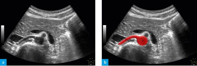
Note the speckling pattern seen across all tissues and the clear differences in tissues between Rectus Abdominis and the surrounding fat.
From Radiology Key
I hope you found this quick whistle stop tour of ultrasound imaging useful and informative. I will be writing some more advanced ultrasound guides as I go through my revision for my upcoming Imaging with Non-Ionising Radiation exam. I will also be creating guides for other imaging modalities as I work through my notes. This year I have undertaken a Bachelor’s degree in Imaging Sciences at King’s College London, read more here.
I always love to connect with my readers. So please do get in touch on Twitter and Linkedin! You can also use the contact form on the website or leave a comment below. You can SUBSCRIBE for updates and post notifications at the bottom of this page.
Thank you for reading!


-
-
6 years
-
Tagged Admissions, Medical School