


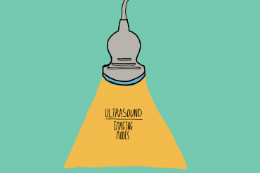
A Medical Student’s Guide To: Ultrasound Imaging Modes
Online Tutoring
I offer online tuition to students studying Biology, Chemistry and Physics at secondary and tertiary levels. I am happy to take on clients from around the world and have a proven track record of top marks achieved by my students. I also tutor for the medical entrance exams, mainly focusing on the UCAT and UCAT ANZ.
To enquire further and set up a free consultation, please use the contact form on this website, drop me an email or contact me through SuperProf.


Ultrasound imaging is a versatile imaging modality, capable of producing a range of images and data graphs. Different modes of ultrasound are used for different purposes, some modes produce clear images of patient anatomy while some provide information on blood flow in real time. Below I’ve briefly explained the most commonly used ultrasound modes. For the basics of ultrasound, see my previous post here.
Ultrasound A-Mode: Amplitude Mode
Amplitude mode ultrasound can be used to create graphs of amplitude against depth. It is the simplest type of ultrasound. A single transducer produces ultrasound waves in a straight line. Destructive therapeutic ultrasound is also A-Mode.
The graphs produced in A-Mode display the amplitude of reflected ultrasound waves reflected at each depth from the ultrasound probes.


Ultrasound B-Mode: Brightness Mode
Brightness Mode imaging is the most recognisable ultrasound imaging technique. Multiple transducers scan a plane through the tissues, displaying a two-dimensional image on a screen. These images express amplitude of received signal as a greyscale intensity.

Ultrasound M-Mode: Motion Mode
Motion mode imaging utilises rapid B-Mode imaging to measure movement of anatomical structures or surgical instruments. The images are displayed in quick succession on a monitor. Anatomy in one line can be isolated and the change in depth tracked against time, as seen below.


Ultrasound Doppler Imaging
Doppler imaging utilises the Doppler effect, the change in frequency caused by the motion of a transmitter relative to a receiver. In the case of ultrasound imaging, where the transmitter and the receiver are the same, the Doppler effect measures the relative movement of the receiving transducer to the reflector of the ultrasound waves.
There will be another article dedicated to the Doppler effect, but in short: the change in frequency is proportional to the velocity of the imaged tissue (such as blood). Therefore, the velocity of the movement can be measured.
Doppler imaging can be combined with B-Mode imaging to create a dual image display: tissue structures displayed in greyscale and tissue velocities displayed in colour.


I hope you found this quick whistle stop tour of ultrasound imaging modalities useful and informative.
I will be writing some more advanced ultrasound guides as I go through my revision for my upcoming Imaging with Non-Ionising Radiation exam. I will also be creating guides for other imaging modalities as I work through my notes. This year I have undertaken a Bachelor’s degree in Imaging Sciences at King’s College London, read more here.
I always love to connect with my readers. So please do get in touch on Twitter and Linkedin! You can also use the contact form on the website or leave a comment below. You can SUBSCRIBE for updates and post notifications at the bottom of this page.
Thank you so much for reading!


Comments